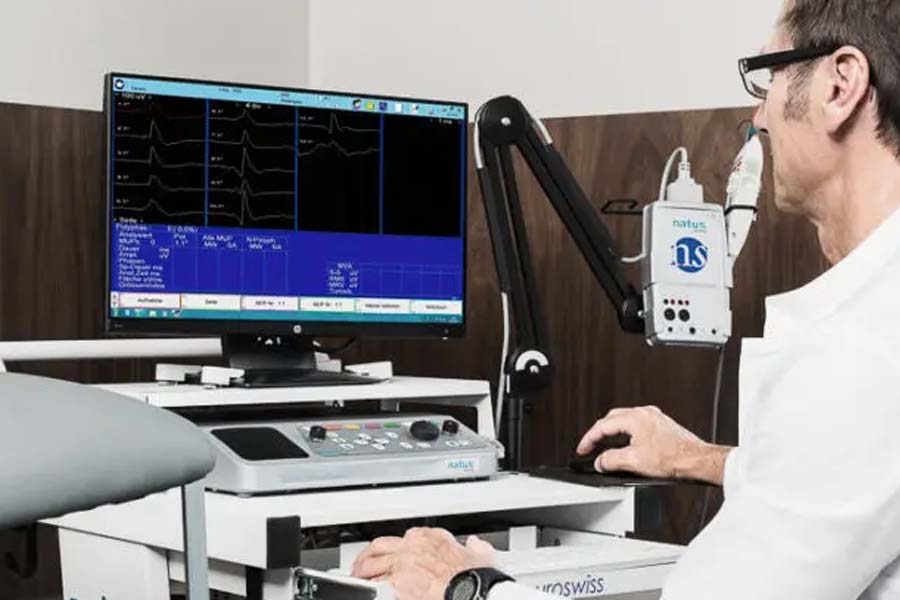
ENMG Test – Electroneuromyography
What is electroneurography (ENG)?
ENG is a test of nerve conduction in peripheral nerves. It measures the speed at which a nerve transmits electrical signals (nerve conduction velocity). In addition, it measures how well an electrical nerve stimulation is transmitted to the corresponding muscle (neuromuscular transmission).
What is electromyography (EMG)?
EMG is a diagnostic procedure that measures the electrical activity of a muscle. It provides information on whether the muscle itself is diseased or the nerve that supplies information to that muscle is not functioning adequately.
How does ENG work?
In motor neurography, the nerve being tested is electrically stimulated at two points. The time from the nerve stimulation to the corresponding muscle contraction is measured. This time is very short – only a few thousandths of a second – and must be determined electronically. The muscle contraction is registered by a computer using surface electrodes.
The nerve is stimulated at two different locations. The nerve conduction velocity (NCV) is calculated from the difference in conduction times and the distance between the two stimulation sites.
How does EMG work?
The doctor disinfects the skin and inserts a very thin needle electrode directly into the patient’s muscle. Amplifiers are used to derive the activity of individual muscle fibers inside a muscle. A computer stores the measured voltage fluctuations and presents them as a signal. These fluctuations can also be heard as noise and crackling through a speaker. Experienced doctors can make an assessment about the nature of an injury based on the sounds alone. The exact analysis is then carried out on the computer. The doctor pays particular attention to:
- Electrical signals that occur immediately after the needle insertion
- The appearance of spontaneous signals in a relaxed muscle (spontaneous activity)
- Signals that occur during gentle muscle contractions in the course of the examination
What is the purpose of ENG?
With the help of an ENG, doctors can classify the type and severity of various peripheral nerve diseases. The following can be diagnosed with this method:
- The severity of a polyneuropathy, which is a disease of the peripheral nerves that can have various causes (including diabetes mellitus or chronic alcohol abuse).
- The exact location and severity of nerve damage caused by injury.
- The extent of compression symptoms of nerves in the area of the legs and arms, such as carpal tunnel syndrome (CTS) at the wrist.
What is the purpose of EMG?
With the help of an EMG, the type and severity of various muscle and nerve diseases can be determined. A thorough neurological examination should always be carried out beforehand in order to establish the suspected diagnosis. Subsequently, the doctor can selectively examine only certain muscles. This is advantageous for patients because the needle EMG is not a pleasant examination.
A relaxed muscle normally does not show any electrical activity. However, even with slight contraction, electrical signals occur, which further increase with stronger muscle movements. Certain diseases of the muscles, peripheral nerves, or nerve roots at the exit from the spine result in abnormal or specific patterns in the electrical activity of the punctured muscle.
The following questions can be answered with an EMG:
- Is a muscle weakness caused by a disease of the muscle or the corresponding nerve?
- In case of muscle paralysis caused by injury or inflammation of the supplying nerve, the EMG provides information about the chances of healing (prognosis). It is important whether there is still residual activity of the muscle or whether there are signs of nerve recovery through regeneration of nerve fibers (so-called reinnervation).
- The exact muscles affected by nerve damage are examined, allowing the location of the nerve damage to be precisely determined. This allows for targeted imaging of the corresponding body region, for example, by computer tomography.
What should be considered prior to ENG?
A comprehensive neurological examination should precede the ENG in order to test as few nerves as possible, as electrical stimulation of the nerves can sometimes be uncomfortable for the patient.
What should be considered prior to EMG?
EMG should not be performed on patients taking blood thinners (e.g. anticoagulants) or with a bleeding disorder. In urgent cases, the doctor may still be able to examine individual, smaller hand or foot muscles, but must weigh the risk of bleeding into the muscle.
Although our laboratory only uses disposable needles, the doctor must be informed of any infectious diseases the patient may have that can be transmitted through blood contact (e.g. hepatitis B or C, or AIDS).
How is ENG performed?
The nerve is electrically stimulated at two points along its course. For nerves that supply the hand muscles, such points can be found, for example, in the upper arm, elbow, or wrist.
Surface electrodes for recording muscle contractions are attached to the finger muscles in this case. During the stimulation, the patient feels an electric sensation.
Two examination results are particularly important:
- Diseases of the myelin sheath, which surrounds the nerves, lead to a slowing of nerve conduction velocity.
- In diseases that affect the interior of the nerves, nerve conduction velocity may be normal, but the height of the electrical nerve response (amplitude of the nerve potential) is lower.
Conclusions about possible nerve damage can also be drawn from the strength of the muscle contraction.
How is EMG performed?
The patient lies as relaxed as possible on an examination table. For needle EMG, the doctor disinfects the skin and inserts a thin needle electrode directly into the muscle. After that, the patient has to wait until the muscle has calmed down after the puncture. Then the patient first contracts the muscle slightly and then fully. The computer records all electrical activities of the muscle.
Possible complications of ENG and EMG
The electrical impulses in electro-neurography are often perceived as unpleasant but tolerable by patients. Serious complications are not known.
Unfortunately, needle EMG is not a painless examination. However, most patients find the puncture pain of the extremely thin needle tolerable. In very rare cases, infections or bruises can occur in the area of the puncture site.
